

Handheld Ultrasound
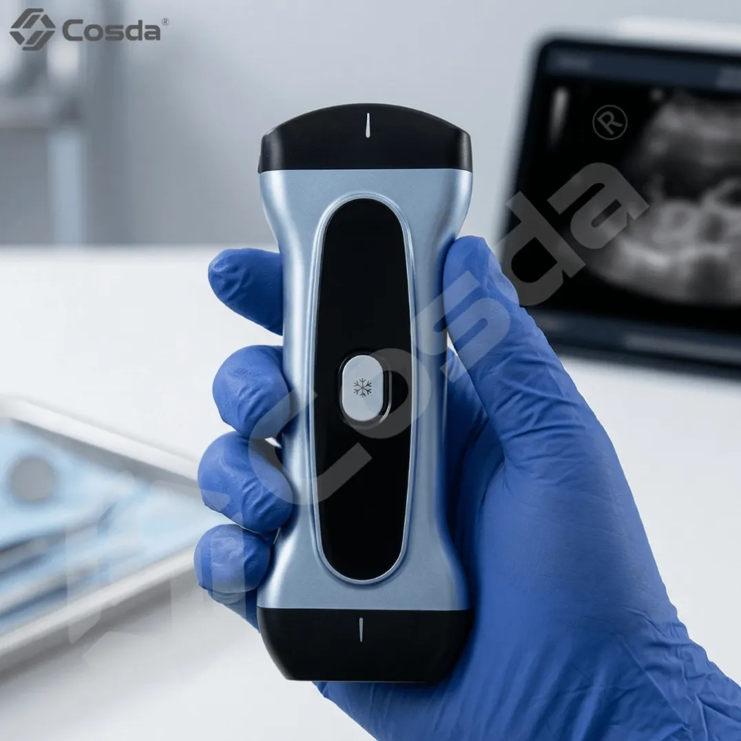
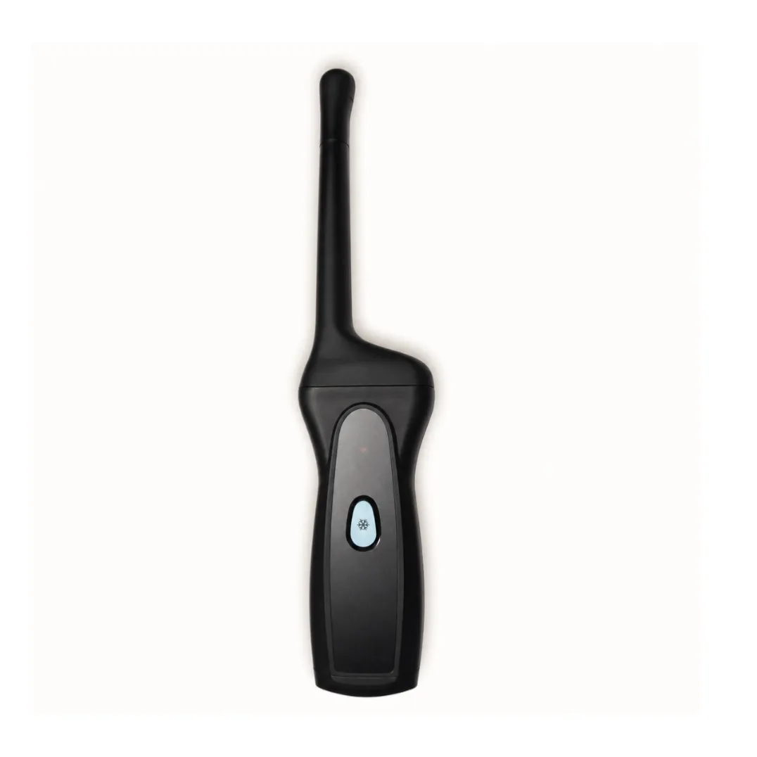
Wireless Color Doppler 3-in-1 Ultrasound Probe | 192-Element Handheld Scanner
Wireless handheld ultrasound with 192 elements: convex, linear and cardiac probes in one device. Color Doppler, panoramic imaging, needle enhancement. Works with iOS, Android and Windows. CE certified, 2-year warranty.
Specifications
- Probe Types
- 3-in-1: Convex / Linear / Cardiac
- Imaging Modes
- B, M, Anatomy M, Color Doppler, PW Doppler, PDI, DPDI, TDI, Color M
- Advanced Features
- Panoramic Imaging, Needle Enhancement, TSI (1540–1620 m/s), Automatic Tracing, Wide Field Imaging
- Frame Rate
- Up to 60 fps
- Compatibility
- iOS, Android, Windows
- Certification
- CE/FDA/ISO
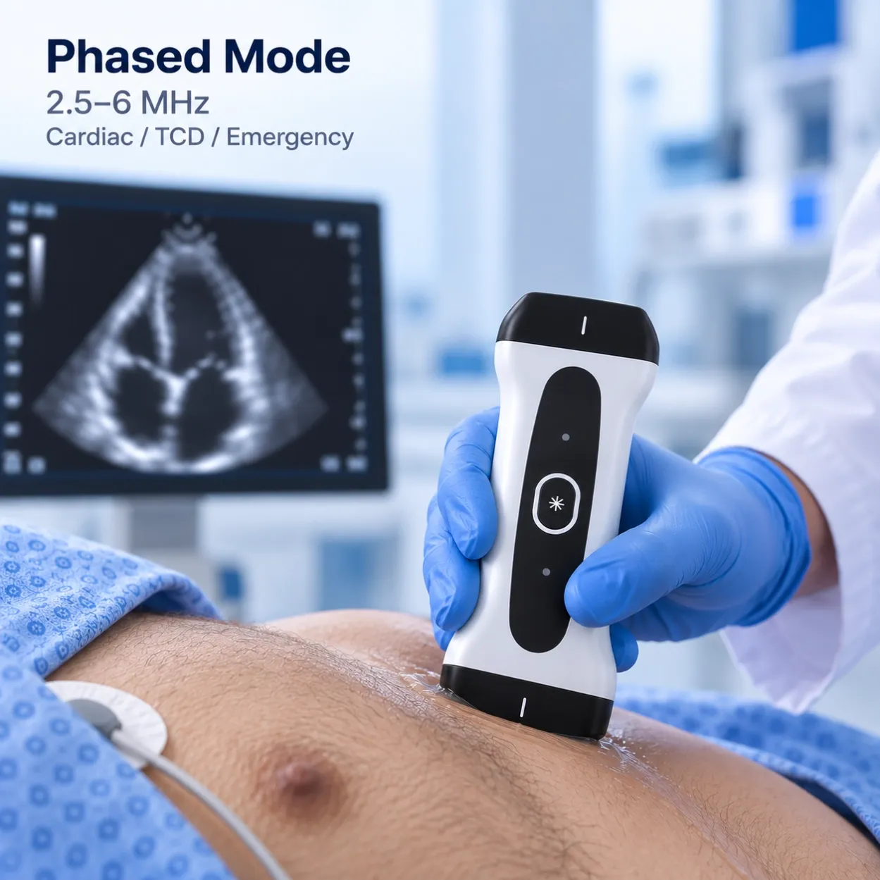
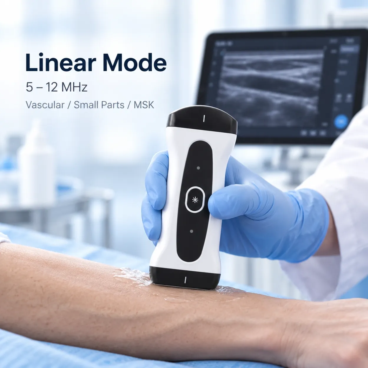
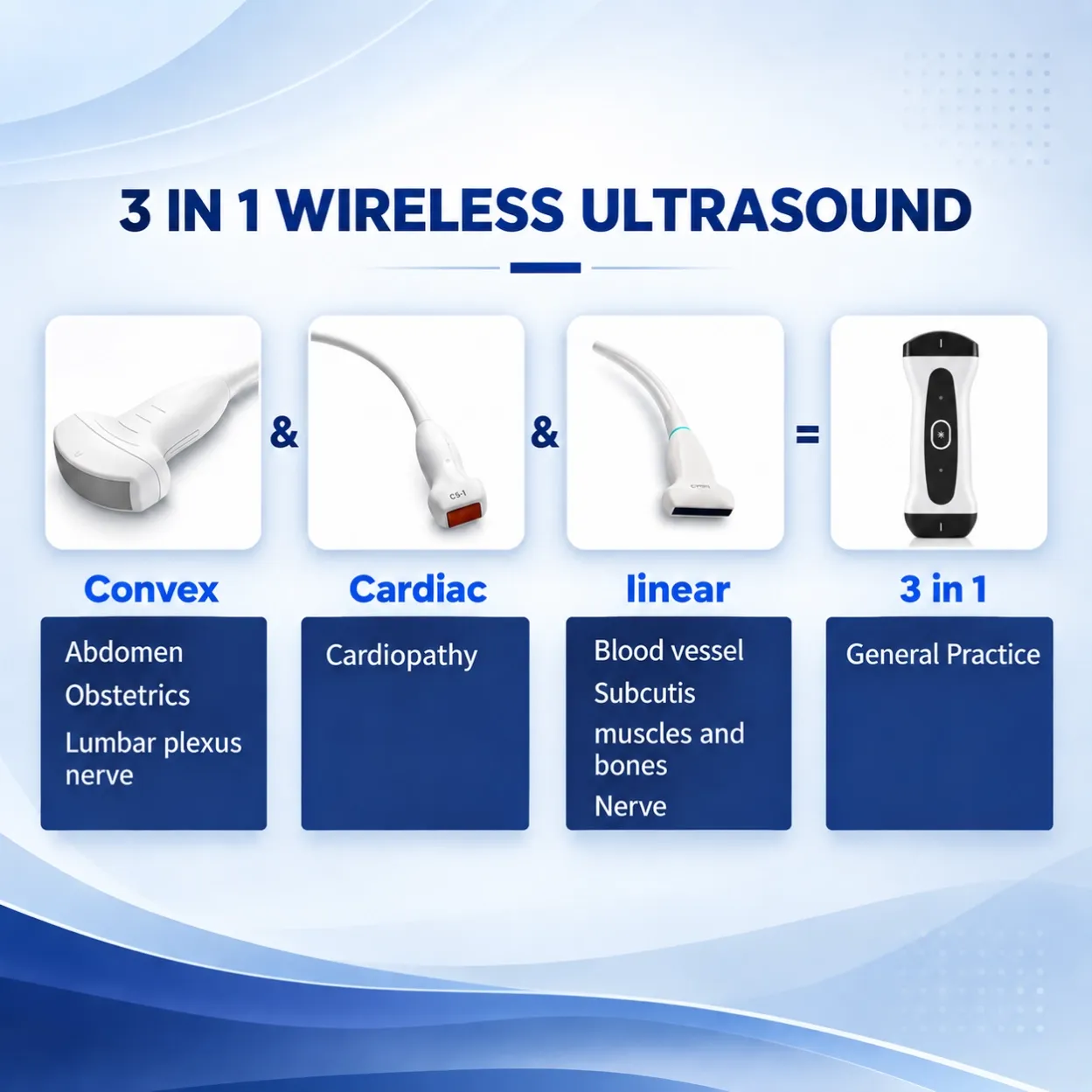 | One Probe, Three Applications (3-in-1) Convex Mode Abdomen · Obstetrics · Lumbar plexus nerve blocks Linear Mode Carotid & peripheral vessels · Thyroid · Nerve blocks (ulnar, median) · MSK / elbow joint Cardiac Mode Cardiac B-mode · Color Doppler · M-mode · Anatomy M-mode No more carrying multiple probes — switch modes in the app and examine any patient with one device. |
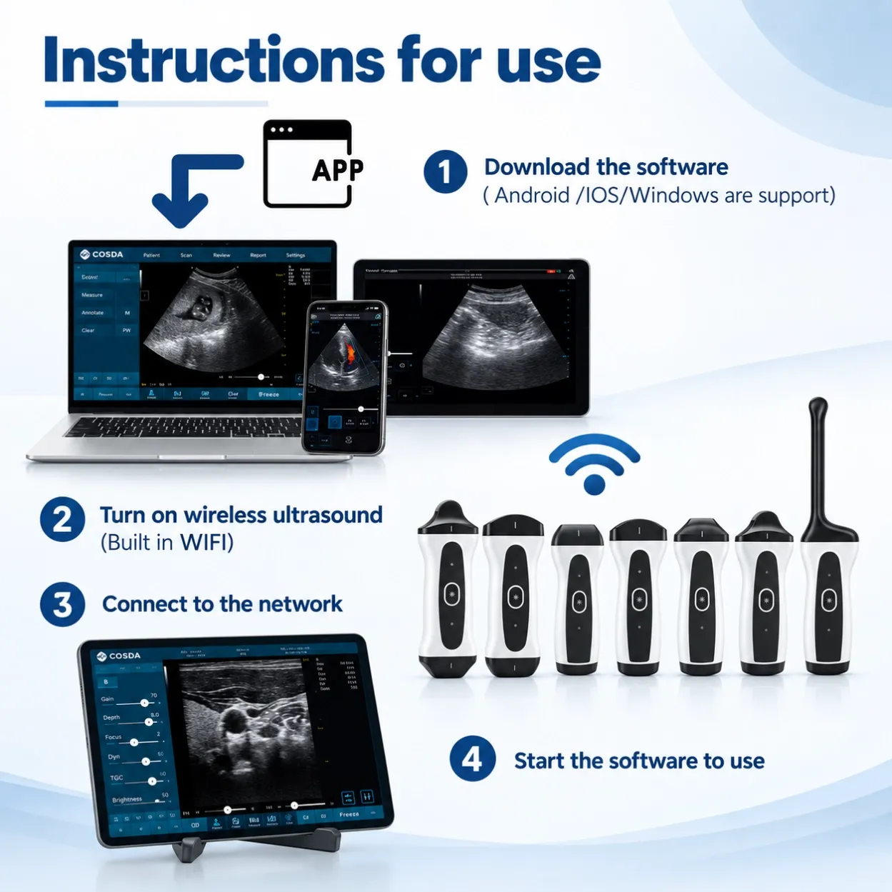
Get Started in 4 Steps
|  |
Advanced Imaging Features
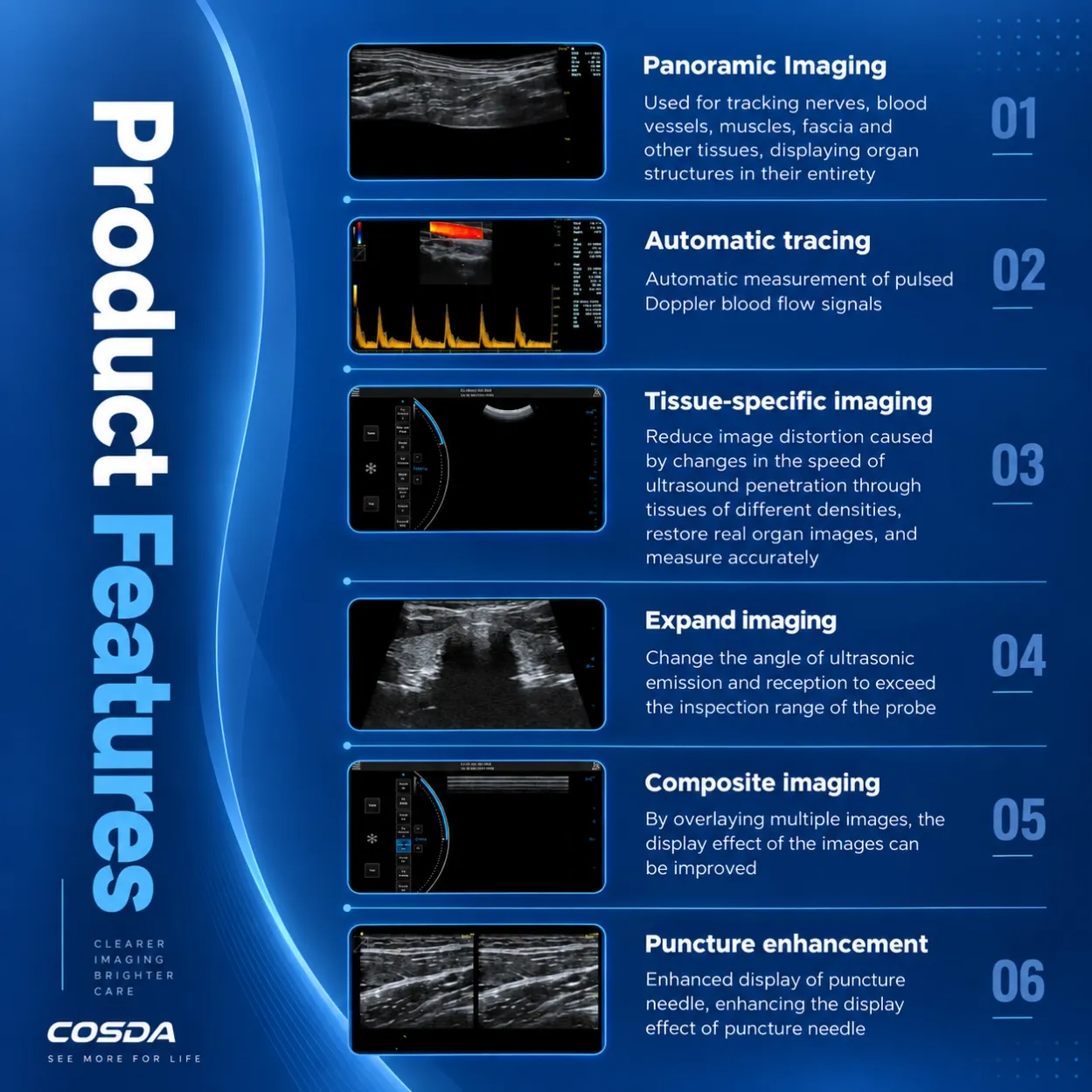
Panoramic Imaging Track nerves, blood vessels, muscles, and fascia; display organ structures in their entirety beyond the probe's physical footprint.
Automatic Tracing Automatic measurement of pulsed-wave Doppler blood flow signals faster, more repeatable measurements.
Tissue-Specific Imaging (TSI) Reduces image distortion caused by varying sound speed through tissues of different densities, restoring true anatomy for accurate measurement.
Extended-Field Imaging Adjusts ultrasound emission/reception angle to exceed the native field of view of the probe.
Compound Imaging Overlays multiple frames to improve image clarity and reduce speckle.
Needle Enhancement Enhanced visualization of the puncture needle for smoother, safer guided procedures.
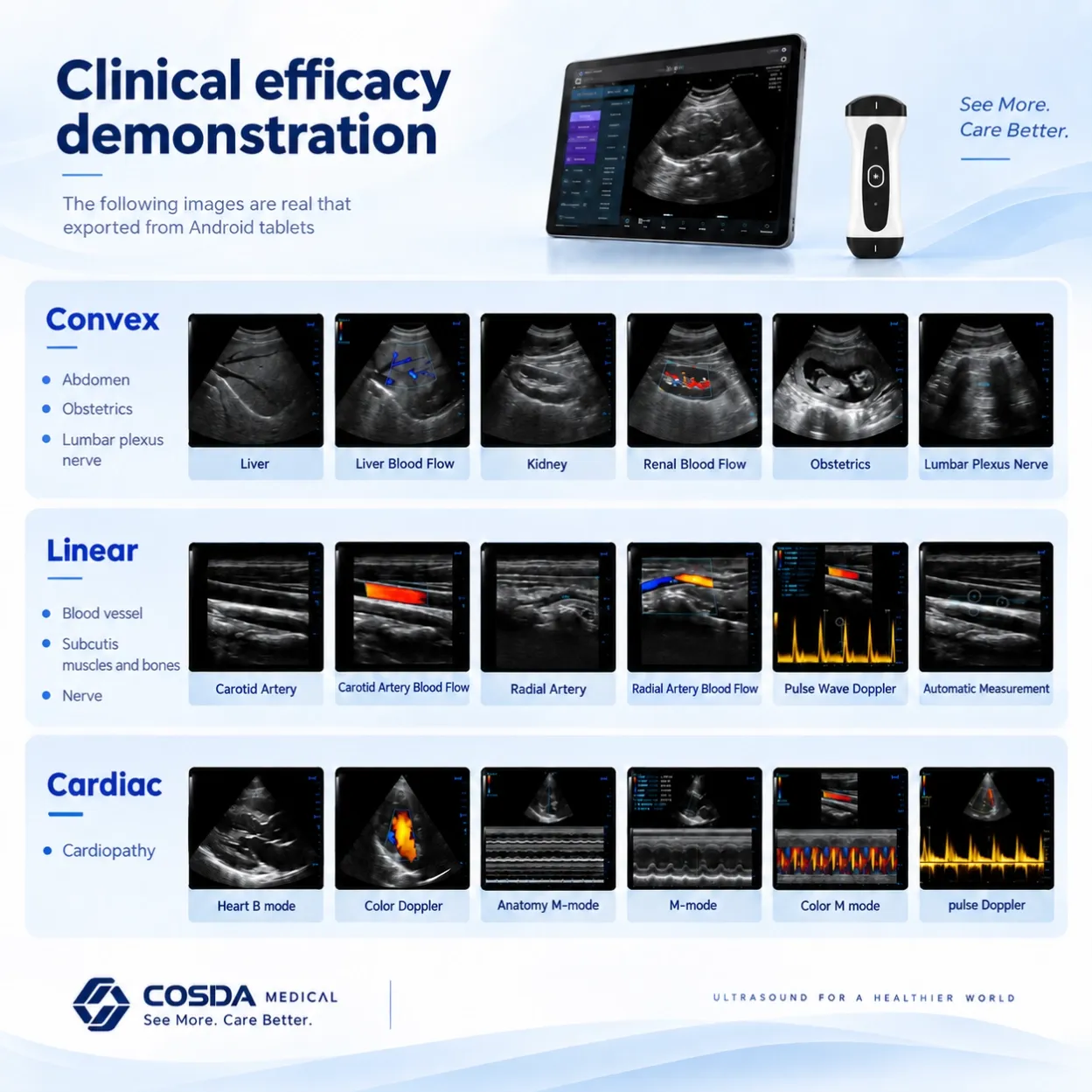
Real Clinical Images
(Place exported sample images here, grouped by probe)
All images exported directly from an Android tablet — no post-processing.
- Convex: Liver · Liver blood flow · Kidney · Renal blood flow · Obstetrics · Lumbar plexus
- Linear: Carotid artery & blood flow · Radial artery & blood flow · Pulse-wave Doppler with auto measurement · Thyroid · Elbow joint · Ulnar & median nerve
- Cardiac: Heart B-mode · Color Doppler · Anatomy M-mode · Color M-mode · Pulse Doppler
3 in 1 Color Doppler Wireless Linear Ultrasound Probe
•Support wireless charging and connection with IOS, Android, Windows
•Frame rate can be up to 60fps
•Support wireless charging and connection with IOS, Android, Windows
•Tissue-specific imaging(TSI):1540m/s--1620m/s
•Freeze image post-processing: After freezing/saving the image, you can measure/annotate it again.
•Display mode:
IOS, Android B/M/CF/PW/PDI/DPDI/Puncture Enhancement/Panorama/Color M
Window B/2B/4B/M/CF/PW/PDI/DPDI/Puncture Enhancement/Panorama/Color M /Anatomy M/TDI/Wide Field Imaging/Contrast Enhanced Mode/Automatic Tracing
• Frequency
Convex 2.5/3.5/4.0/5.0/6.0/H3.6/H4.0/H5.0/H6.0
Linear 5.0/7.5/9.0/10.5/12/H10/H12
Cardiac 2.5/3.5/4.0/5.0/6.0/H3.6/H4.0/iH3.6/iH4.0
•Professional department software data package: Abdomen,Obstetrics,Gynecology,Cradiac,Vascular,Small organs,Urology,Spine,Pediatrics,Cranium
FAQ
Q: Which devices does the PAU2A work with?
A: Any iOS or Android phone/tablet and Windows PC. Just download the free app and connect via the probe's built-in Wi-Fi. No internet required.
Q: How long does the battery last?
A: Around 2 hours of continuous scanning on a full charge — enough for a typical clinic session or rounds.
Q: Does it support color Doppler?
A: Yes — B-mode, M-mode, Anatomy M-mode, color Doppler, and pulsed-wave Doppler with automatic measurement.
Q: Can one probe really replace three?
A: Convex, linear, and cardiac arrays in a single handheld device. You select the mode in the app, so one probe covers abdomen, OB, vascular, nerve, MSK, and cardiac exams.
Q: Is it suitable for needle-guided procedures?
A: Yes. Needle Enhancement mode improves needle visibility, and Panoramic Imaging helps track nerves and vessels during blocks.