Sep 5, 2026Buying Guides
Equine Ultrasound Machine: Selection Guide for Vets
What equine practice demands from an ultrasound machine: linear frequencies for tendon work, wireless field logistics, battery life, and a selection checklist.
An equine ultrasound machine lives a harder life than its clinic-bound cousins. It gets carried to stalls and paddocks, used around a half-ton animal that cannot be asked to hold still, and expected to resolve fine tendon fibers at depth — often on battery, in sunlight, away from the clinic. So choosing one is less about finding the "best" machine and more about matching three things to equine work: the right probe frequencies for tendon and soft tissue, logistics that survive the farm, and a workflow that follows the horse.
If you are an equine practitioner or run a mobile equine practice doing lameness, pre-purchase, or reproductive work, this guide is for you. If your caseload is small-animal only, or you are equipping a referral hospital with fixed imaging suites, a cart-based system conversation is the one worth having instead.
Why Equine Ultrasound Purchases Go Wrong
- The mobile equine vet buys a console that performs beautifully in the clinic demo room and proves exhausting to carry across a farm.
- The default probe that ships with a budget machine covers abdominal work but lacks the high linear frequencies that tendon lameness exams actually need.
- Cables around a nervous horse create exactly the kind of sudden-pull accident that ends probes early.
- The practice re-books exams because images from the field cannot be reviewed or sent to the referring surgeon the same day.
- Clients start comparing your delays to the practice down the road, and the equipment becomes the reason the schedule slips.
- Every one of these failure points is checkable before purchase — the checklist at the end covers them.
What Equine Work Actually Demands
Equine ultrasound splits into three job families, and each one pulls on different hardware:
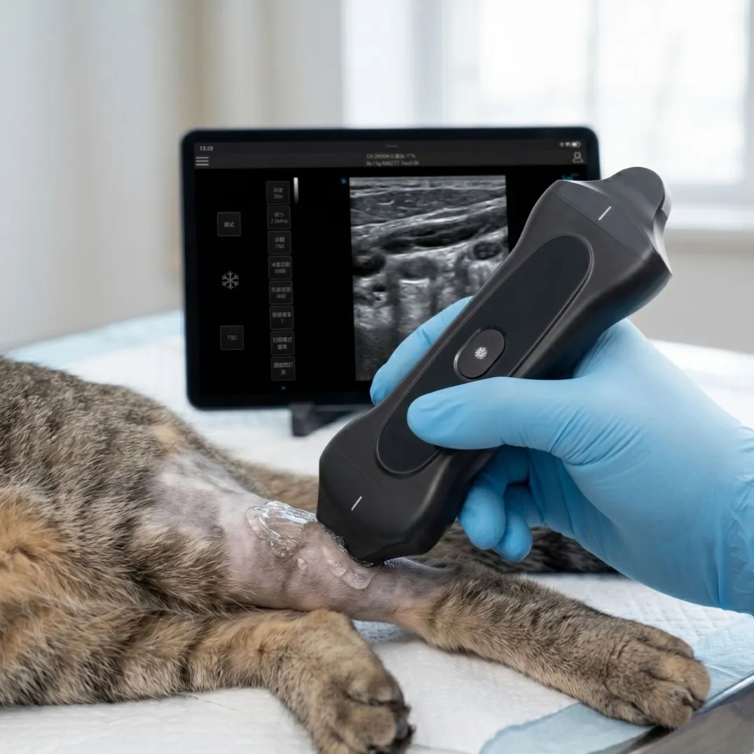
Tendon and ligament (lameness) work is the signature equine exam. Tendon fibers are best resolved with high-frequency linear arrays — in equine practice this generally means scanning in roughly the 7.5–12 MHz range, where fine fiber architecture is visible. This is industry-standard practice, not a vendor claim: the linear band is where lameness work lives. Our wireless linear probe publishes a 5.0–12.0 MHz band (5.0/7.5/9.0/10.5/12 MHz, plus harmonic modes H10/H12), which covers that clinical range.
Abdominal and thoracic work needs depth, which is convex territory. Our 3-in-1 wireless probe carries a convex band (2.5–6.0 MHz) alongside the linear and a phased array, so one pocket probe handles a belly scan and then switches to tendon work without a probe change — useful when the horse is not enthusiastic about waiting.
Reproductive work is where honesty matters most. Transrectal examination of the reproductive tract has specific probe and technique requirements, and we will not tell you a handheld probe replaces a purpose-built rectal setup — that depends on your protocol and the exam. What the wireless format does change is the rest of the logistics: no cables in the stocks, images on a tablet you already carry, and exams documented the moment they happen. Ask any supplier — including us — to be explicit about which reproductive protocols their hardware supports.
The Wireless Question: Gimmick or Field Upgrade?
Around horses, cables are a safety problem, not just a convenience problem. A spooked horse and a tethered probe meet exactly once. A wireless probe that streams to iOS, Android, or Windows over Wi-Fi removes the cable entirely, and wireless charging means there is no charging port to fill with arena dust.
The practical checklist for the wireless claim, beyond the absence of cables:
- Battery that survives a farm round. Our handheld probes are rated around two hours per charge with fast charging (about 100% in 60 minutes) — enough for a typical ambulatory round, but do the math on your own day.
- Image handling on the device you already own. Images land on a tablet or phone you carry anyway, and can be shared with the referring vet the same day. That closes the "field exam to same-day referral" gap that cable-bound setups often leave open.
- Modes where you need them. The 3-in-1 publishes B/M/CF/PW/PDI/DPDI across its three transducers, so Doppler questions do not require a second machine.
Clinic Base, Field Kit, or Both?
Practices split into two realistic patterns. Mobile-first practices lean on a handheld probe as the primary scanner and keep clinic-based imaging minimal. Clinic-anchored practices doing mixed caseloads often pair a laptop-style console — our laptop portable color Doppler unit runs 3–4 hours on battery, supports ten-plus probe types, and boots in about 12 seconds — with a handheld probe for farm calls. The existing guide to essential veterinary ultrasound features covers the veterinary-specific selection criteria in depth.
Matching the Modes: What B, CF, PW and PDI Do on the Horse
Spec sheets list modes as acronyms, and it is worth being concrete about what each one does on a horse:
- B-mode is the structural image: tendon fibers, uterine horn, lung surface. Everything starts here.
- M-mode tracks motion along a single line — useful for cardiac questions on the foal or the performance horse.
- Color flow (CF) and power Doppler (PDI) show blood flow where it happens. In tendon work, new vessel formation in the lesion is part of how healing is monitored between exams; in the abdomen, they answer perfusion questions.
- PW Doppler adds velocity measurement to a vessel you have already found with color.
Two practical notes for equine buyers. First, mode support can vary with the connected device — our probe publishes the full mode set, and we tell distributors to verify their exact tablet runs every mode they need, on every ecosystem they carry. Second, the difference between PDI and DPDI (directional power Doppler) matters in vascular questions; if your caseload includes those, ask which one the vendor's "power Doppler" actually means.
Three Questions Equine Buyers Ask Before Ordering
Can one probe really cover both tendon and abdominal exams? It depends on the probe. A single-frequency linear probe covers tendon work but struggles with abdominal depth. This is exactly why the 3-in-1 exists: convex (2.5–6.0 MHz), linear (5.0–12.0 MHz), and phased array in one unit, switched in software, so a farm round does not mean a bag of probes. The trade-off is explicit: the 3-in-1 trades a small amount of per-transducer optimization for the ability to carry one probe instead of three.
How much battery does a farm round actually need? Add up scan time, not appointment time — a probe that idles between exams still drains slowly. Our handhelds are rated around two hours of active use per charge, and the wireless charging scheme means a probe can top up in the truck between calls (about 43% in 30 minutes). Practices running all-day ambulatory schedules typically carry a charged second unit or an in-cab charging pad; price that into the comparison rather than discovering it in the field.
Do the images work with the software my clinic already runs? The probe streams to iOS, Android, and Windows devices, and images can be exported and shared from the device you scanned on — which closes the field-to-referral gap. What a probe does not replace is a clinic-wide PACS integration; if your referring surgeons want DICOM into hospital systems, that is the laptop console's job. We would rather draw this line clearly than have a buyer discover it after delivery.
A Six-Line Selection Checklist for Equine Practice
- Confirm the linear band reaches the frequencies your tendon work needs — ask for the published numbers, not "high frequency."
- Count the battery minutes in a real ambulatory round, then double the margin you think you need.
- Handle the wireless workflow end to end: scan, save, annotate, and send to a referrer from the field.
- Ask directly what reproductive protocols the hardware supports, and get the answer in writing.
- Check the modes list (CF/PW/PDI at minimum) if you do vascular or cardiac questions.
If the checklist points you toward a wireless probe, the product pages for the 3-in-1 wireless probe and the wireless linear probe publish the full specification sheets — and if you tell us your caseload mix, we will tell you honestly whether a handheld is enough or whether your practice needs the laptop console behind it.